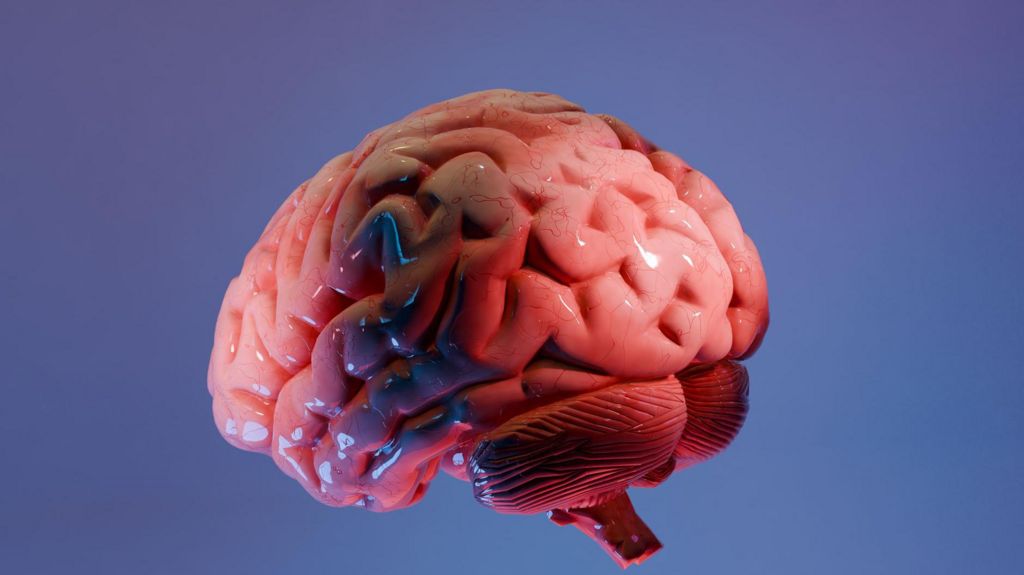
For over 100 years, neuroscience has progressed like early geographic exploration: researchers have pieced together fragments of the human brain’s complex structure from scattered, isolated observations, leaving vast uncharted territory still waiting to be explored. Even today, when clinicians diagnose neurodegenerative conditions such as Alzheimer’s disease, they typically only examine a tiny handful of tissue samples from an organ that contains roughly 86 billion neurons – meaning most of the brain’s landscape remains unseen.
Now, a team of scientists at the Sudha Gopalakrishnan Brain Centre (SGBC) based at the Indian Institute of Technology, Madras (IIT-M) have announced a breakthrough that addresses one of the largest gaps in modern neuroscience. The team has created what they call the world’s most detailed three-dimensional cellular-resolution atlas of the human brainstem, a fully digital mapping tool that allows researchers to navigate seamlessly from full-brain MRI scans all the way down to the level of individual nerve cells.
Named Anchor, short for *Atlas of Neurochemical Characterisation of the Human Brainstem with 3D Reconstruction*, the new resource integrates data from more than 500 tissue samples harvested from foetal, childhood, and adult human brains. Unlike high-resolution brain mapping projects that rely on expensive molecular sequencing techniques, Anchor is constructed from high-magnification microscope images of thin tissue slices, enabling it to generate a comprehensive 3D map of the brainstem that identifies more than 200 distinct cell clusters and neural pathways. Eight targeted chemical markers are used to differentiate between cell types, producing one of the clearest portraits ever compiled of this critical but vastly understudied brain region.
Though the brainstem makes up only a tiny fraction of the brain’s total volume, it is indispensable to human survival. Serving as the physical and functional link between the cerebral cortex and the spinal cord, the brainstem regulates core involuntary processes that sustain life: breathing, heart rate, sleep-wake cycles, and basic movement control. Even small lesions to tiny cell clusters within the brainstem can cause catastrophic, fatal damage, but the region’s tightly packed, intricate architecture has long stymied efforts to map it in fine detail.
The true significance of Anchor extends far beyond adding another anatomical reference to neuroscience. For decades, two core branches of brain research have operated largely in isolation: whole-brain medical imaging, which provides a broad overview of brain structure but lacks fine cellular detail, and cellular pathology, which examines individual cells one slice at a time but cannot contextualize those observations within the brain’s overall 3D architecture. Anchor bridges this long-standing divide.
Shubha Tole, a leading neuroscientist at the Tata Institute of Fundamental Research, described the project as a visionary achievement that places India at the forefront of global neuroscience, calling it an unprecedented integration of engineering, neuroscience, and clinical medicine.
Rebecca Folkerth, a neuropathologist affiliated with Harvard Medical School and New York University who collaborated on the project, explained the long-standing limitations of traditional brain research practices. “As a neuropathologist, I begin by examining an entire brain with the naked eye before looking at small pieces under the microscope,” she said. “For Alzheimer’s disease, we may examine only 15 to 20 sections – just a fraction of a percent of the whole organ.” This fragmented approach has been the standard since the pioneering work of Santiago Ramón y Cajal, the father of modern neuroscience, more than a century ago. Modern MRI can capture the full structure of a living brain, but cannot resolve individual cells; microscopes can show individual neurons, but only in disconnected, thin tissue slices.
“What the Indian centre has created is essentially what I dreamed of early in my career – to have brain scans match the brain’s microscopic anatomy,” said Folkerth, who has examined thousands of human brains over a 30-year career in research.
Anchor’s interactive design allows users to zoom continuously from a full-resolution MRI view of the entire brainstem down to individual neurons, while preserving the precise spatial relationships between all structures. To advance global research, the SGBC team has made the full atlas freely accessible online, with the goal of it becoming a standard reference tool for neuroscientists, neurologists, and neurosurgeons around the world.
The potential applications of the atlas reach far beyond basic anatomical research. By comparing healthy mapped brainstem tissue to samples from patients with neurological conditions, researchers can gain new insights into a wide range of disorders including Parkinson’s disease, stroke, Alzheimer’s disease, and sudden infant death syndrome (SIDS). A more accurate 3D map of the brainstem will also allow neurosurgeons to navigate this delicate, high-stakes region with greater precision and lower risk during procedures.
Importantly, Anchor is not designed to be a direct clinical diagnostic tool. Instead, its greatest value lies in enabling researchers to ask and answer new questions that were previously impossible to address. Partha Mitra, a leading brain scientist at Cold Spring Harbor Laboratory in New York who has collaborated with SGBC, notes that detailed cell-level atlases like Anchor could have a transformative impact on neurological disease research. By comparing cell-by-cell structure between healthy brains and those affected by conditions such as Alzheimer’s or autism, researchers can identify previously unrecognized pathological changes. Mitra also added that the atlas could help unpack how viral infections such as Covid-19 trigger long-term neurological damage in patients suffering from long Covid.
Using stroke as an example, Folkerth explained that the new atlas has already revealed previously unrecognized anatomical features that could help clinicians preserve brain tissue that is injured but not yet permanently damaged, ultimately improving patient outcomes. The resource’s accessible design also makes large-scale brain mapping far more affordable for research institutions around the world, as it relies on standard high-resolution microscopy rather than costly molecular techniques. This affordability allowed the SGBC team to map the human brainstem at an unprecedented scale that would have been out of reach for many projects using alternative approaches.
The project reflects a broader shift in modern neuroscience, where progress now depends as much on advances in engineering and computational science as it does on biological research. Around 20 SGBC scientists spent 18 months manually analyzing more than 200 brain sections, integrating MRI data, microscopic anatomy, and 3D reconstruction into a single unified digital resource. Today, the SGBC brings together more than 200 researchers, engineers, and technicians working with international collaborators across the globe to advance human brain mapping.
Mohanasankar Sivaprakasam, head of SGBC, noted that while the brains of several common animal model species have been mapped in extraordinary detail, the human brain remains vastly under-charted due to the limited availability of well-preserved human tissue for detailed research. While many brain atlases already exist, each fills a unique niche: MRI-based atlases capture broad structure but not cellular detail, histological atlases offer cellular resolution but lack 3D context, and newer molecular atlases identify cell types but do not yet map large-scale 3D architecture at scale. Anchor fills a critical gap that no existing resource has addressed.
Even with this breakthrough, much remains unknown about the human brain: researchers still have very little understanding of how the brain’s roughly 20,000 proteins are distributed across different regions and cell types, a frontier that will define the next generation of brain mapping research. As Folkerth puts it, “Every brain is a treasure chest of new knowledge.”
Looking ahead, SGBC plans to continue its work by imaging more than 100 whole human brains spanning different life stages and neurological conditions, including Alzheimer’s disease and dementia, to build a comprehensive reference library that will reveal how disease reshapes the human brain one cell at a time. While the new Anchor atlas will not solve all the mysteries of the human brain overnight, it gives researchers a far more detailed map to guide their work, helping them ask better questions – and ultimately find the answers that could transform neurological care.